Anterior Cruciate Ligament (ACL)  | Meniscal Injuries | Medial Collateral Ligament (MCL) Injury | Posterior Cruciate Ligament (PCL) Injuries | Lateral Collateral Ligament (Lcl) Injuries  | Patellar Dislocation | Patellar Tendinopathy | Iliotibial Band Syndrome (ITBS)  | Osgood-Schlatter Syndrome | Patellofemoral Pain Syndrome (PFPS)  | Knee Osteoarthritis (Oa)

Anterior Cruciate Ligament(ACL)

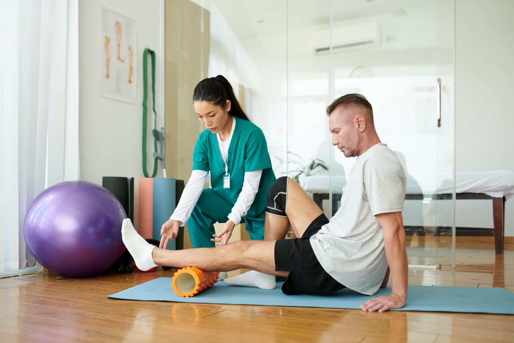
The anterior cruciate ligament (ACL) is one of the ligaments in the knee which provide stability to the knee joint. ACL injuries commonly occur in sports which require rapid deceleration, quick changes of direction, and jumping or landing such as netball, soccer and football. The severity of the injury can range from a mild tear to a complete rupture. ACL injuries are more common in females than males due to differences in anatomy between male and female knees. They most commonly occur in females from 15-19 years old and males from 20-24 years old.  

Symptoms:

  • Sudden onset usually during sport 
  • A loud ‘pop’ or popping sensation in the knee
  • Feeling of instability or giving way with weightbearing 
  • Rapid swelling 
  • Inability to weight bear in more severe cases

What might a treatment plan look like?

Typical recovery following an ACL repair is 9-12 months.  This plan may vary based on your individual situation, your level of injury, whether or not surgery is required, and the goals you want to achieve.  An example of a treatment plan is: 

  • Physiotherapy once or twice per week for 3-4 weeks, then fortnightly sessions for 4 sessions, monthly visits for another 3-4 months, then visits every 6 weeks for another 3-6 months. 
  • Hydrotherapy provided by a physiotherapist or exercise physiologist in the early rehabilitation stages may be beneficial
  • The later stages of your treatment plan may be provided by an exercise physiologist, and may be targeted at sports specific programming if required.

Which of our team members work with ACL injuries? 

  • All of our physiotherapists
  • All of our exercise physiologists

Meniscal Injuries

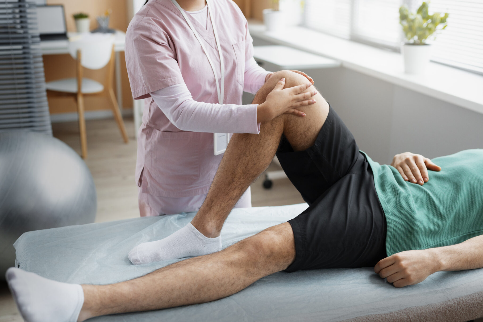
The meniscus is a C-shaped piece of cartilage which sits in the knee between the thigh bone and the shin bone. There are two menisci in each knee. They are responsible for helping to provide shock absorption and stability to the knee. Meniscal injuries can occur during activities that put pressure on the knee and rotate the knee joint. Meniscal injuries can occur in younger populations or the older population where degenerative meniscal tears are more common. 

Symptoms 

  • Pain, especially when the area is touched
  • Swelling 
  • Feeling of the knee locking, catching or giving way 
  • Inability to move knee through full range of motion 

What might a treatment plan look like? 

This can vary depending on whether or not surgery is required and your individual goals. Conservative treatment will likely take around 6-8 weeks, while rehabilitation after surgery may take up to 3 months. An example of a treatment plan is: 

  • Physiotherapy once or twice per week for 2-3 weeks, then fortnightly sessions for 3-4 weeks. 
  • Hydrotherapy under the supervision of a physiotherapist or exercise physiologist is helpful in managing Meniscal injuries.
  • Moving to monthly physiotherapy and/or exercise physiology for limb strengthening and/or sports specific training.

Which of our team members work with meniscal injuries?

  • All of our physiotherapists 
  • All of our exercise physiologists

Medial Collateral Ligament (MCL) Injury

The MCL is a ligament which is on the inner side of the knee joint. It is one of the four major ligaments which are responsible for stability of the knee. Meniscal injuries can range from a minor tear to a complete rupture. MCL injuries generally occur in a sporting population and have an obvious cause of injury. 

Symptoms 

  • Swelling 
  • Tenderness over the inside joint of the knee
  • A feeling of instability 

What might a treatment plan look like? 

This will vary depending on the severity of your injury, and your individual goals. Generally MCL injuries recover within 6-12 weeks. An example of a treatment plan might look like this:

  • Phase 1 – (1-4 weeks)   Physiotherapy once or twice a week  
  • Phase 2 (4-8 weeks) – includes increased range of motion and initial strengthening back to functional level  – Physiotherapy once a week.
  • Phase 3 (8-12 weeks) – progression in strength and range to previous sport specific levels.  Exercise Physiology – 1-2 times a week.
  • Hydrotherapy under the supervision of a physiotherapist or exercise physiologist may also be beneficial in the management of MCL injuries.

Which of our team members work with MCL injuries? 

  • All of our physiotherapists 
  • All of our exercise physiologists 

Posterior Cruciate Ligament (PCL) Injuries

The posterior cruciate ligament is one of the four main ligaments in the knee which function as stabilisers of the knee joint. The PCL specifically prevents the shin bone from sliding backwards behind the thigh bone, and assists in preventing excessive rotation of the knee joint. 

A PCL injury typically presents with other knee injuries, including ACL, MCL, posterolateral corner (PLC), or meniscal injuries. 

Symptoms 

  • Pain in the back of the knee
  • Pain when kneeling 
  • Usually minimal swelling

What might a treatment plan look like? 

This will vary depending on the severity of your injury, and your individual goals. Generally PCL injuries recover within 6-12 weeks. An example of a treatment plan might look like this:

  • Phase 1 – (1-4 weeks)   Physiotherapy once or twice a week  
  • Phase 2 (4-8 weeks) – includes increased range of motion and initial strengthening back to functional level  – Physiotherapy once a week.
  • Phase 3 (8-12 weeks) – progression in strength and range to previous sport specific levels.  Exercise Physiology – 1-2 times a week.
  • Hydrotherapy under the supervision of a physiotherapist or exercise physiologist may also be beneficial in the management of PCL injuries.

Which of our team members work with PCL injuries? 

  • All of our physiotherapists 
  • All of our exercise physiologists

Lateral Collateral Ligament (Lcl) Injuries 

The lateral collateral ligament is one of the four main ligaments in the knee. It is on the outer side of the knee joint and it helps to stabilise the knee. LCL injuries are less common than MCL injuries and usually require more force to injure. It is common for LCL injuries to include injuries of the posterolateral corner (the ligaments at the back and outer side of the knee). 

Symptoms 

  • Pain 
  • Side-to-side instability with knee straight 
  • Increased difficulty walking on uneven ground or up and down stairs
  • Swelling

What might a treatment plan look like? 

This will vary depending on the severity of your injury, and your individual goals. Generally people recover from LCL injuries within 6-12 weeks. An example of a treatment plan might look like this:

  • Phase 1 – (1-4 weeks)   Physiotherapy once or twice a week  
  • Phase 2 (4-8 weeks) – includes increased range of motion and initial strengthening back to functional level  – Physiotherapy once a week.
  • Phase 3 (8-12 weeks) – progression in strength and range to previous sport specific levels.  Exercise Physiology – 1-2 times a week.
  • Hydrotherapy under the supervision of a physiotherapist or exercise physiologist may also be beneficial in the management of LCL injuries.

Which of our team members work with LCL injuries? 

  • All of our physiotherapists 
  • All of our exercise physiologists

Patellar Dislocation 

The patella (kneecap) is a bone which sits in front of the knee joint. The tendon of the quadriceps muscle runs over the top of the kneecap and inserts into the shin bone. The main stabilisers of the kneecap are the medial patellofemoral ligament (on the inner side of the kneecap) and the inside portion of the quadriceps muscle. Patellar dislocation occurs when the kneecap is displaced towards the outside edge of the knee. 

Symptoms 

  • Pain 
  • Swelling 
  • Sensation of slipping or ‘popping out’ 
  • Tenderness over inside edge of kneecap 

What might a treatment plan look like?

This can vary depending on the severity of your injury, your clinical findings, and individual goals. Usually recovery from patellar dislocation can take 8-12 weeks. An example of a treatment plan is:

  • Physiotherapy once or twice a week for 2-3 weeks, followed by fortnightly visits for 2-4 sessions. 
  • Prescription of an appropriate and properly fitted brace to support the affected area.
  • Progression to monthly physiotherapy or exercise physiology for 1-2 sessions if further rehabilitation is required.
  • Hydrotherapy under the supervision of a physiotherapist or exercise physiologist may be beneficial 

Which of our team members work with patella dislocation?

  • All of our physiotherapists 
  • All of our exercise physiologists 

Patellar Tendinopathy

The patellar tendon runs from the quadriceps muscle over the kneecap and inserts into the top of the shin bone. Tendinopathies are usually a result of repetitive overloading and commonly occur in jumping sports. When the load applied to the tendon is too great, the tendon can become stressed. If well managed this can heal quickly however if load is continually applied the tendon can become worse. 

Symptoms 

  • Pain directly under kneecap
  • Pain with loading tendon (jumping, changing direction, decelerating) 
  • Usually, no pain at rest 
  • Pain may improve with repeated loading  

What might a treatment plan look like?

This will vary depending on the severity of your pain, clinical findings, and individual goals. 

Patellar tendinopathy can take 8-12 weeks for reduction in pain, and improvement in function, however some people may have ongoing symptoms for 6+ months. A treatment plan might look like:

  • One or two physiotherapy sessions per week for 2 weeks, then fortnightly sessions for 4-5 weeks.
  • Progression to monthly sessions with a physiotherapist or exercise physiologist if ongoing rehabilitation is required.
  • Hydrotherapy under the supervision of a physiotherapist or exercise physiologist may be beneficial 

Which of our team members works with patellar tendinopathy?

  • All of our physiotherapists 
  • All of our exercise physiologists 

Iliotibial Band Syndrome (ITBS) 

The ITB is a band of connective tissue which starts at the outside of the hip and inserts around the side of the knee.  ITBS is usually an overuse injury which causes pain on the outside of the knee. It is more commonly seen in long distance runners and cyclists, and endurance athletes. 

Symptoms

  • Pain on the outside of the knee
  • Pain aggravated by running or cycling 
  • Pain which often develops around the same time/distance during an activity 
  • Longer training sessions or downhill courses can be aggravating

What might a treatment plan look like?

This will vary depending on the severity of your symptoms, clinical findings and individual goals but ITBS rehabilitation typically takes 6-8 weeks. An example of a treatment plan might be:

  • Physiotherapy once a week for 2 weeks, followed by fortnightly visits for 4-6 weeks 
  • Progression to exercise physiology or physiotherapy every 3-4 weeks if required for your individual goals 

Which of our team members works with ITBS?

  • All of our physiotherapists 
  • All of our exercise physiologists 

Osgood-Schlatter Syndrome

Osgood-schlatter syndrome is an overuse injury that occurs in children. It involves inflammation at the tibial tuberosity, the insertion point of the patella tendon at the front and top of the shinbone (just under the knee). The condition is most common during growth spurts when bones, muscles and tendons are growing rapidly. 

Symptoms

  • Pain localised to the front of the shinbone
  • Pain which worsens with activities such as running, jumping and walking up and down stairs

What might a treatment plan look like?

This will vary depending on the severity of your condition, clinical findings, and your individual goals. The length of time required to recover from Osgood-Schlatter syndrome is different for each person and you may have flare-ups of pain on and off until you finish growing. Some cases of Osgood-Schlatter syndrome can take 12+ months to completely resolve, depending on the severity, however you will not necessarily need to see a physiotherapist or exercise physiologist for this whole time. An example of a treatment plan is:

  • Physiotherapy sessions once or twice a week for 2 weeks, progressing to fortnightly for 3-4 sessions. 
  • Progression to exercise physiology every 6-8 weeks, for ongoing strengthening and sport-specific exercises 
  • Physiotherapy as required to manage flare-ups and assist with training modification 

Which of our team members work with Osgood-Schlatter syndrome?

  • All of our physiotherapists 
  • All of our exercise physiologists 

Patellofemoral Pain Syndrome (PFPS) 

Patellofemoral pain is the term used to describe pain in and around the kneecap. This can occur for several reasons including overloading the knee through higher training volumes or intensities, or poor patellar tracking (movement of the kneecap). Loaded activities that are performed with the knee bent can subject the patellofemoral joint to loads ranging from 0.5 times body weight to seven to eight times body weight (eg. when climbing stairs). 

Symptoms

  • Diffuse pain around or under kneecap
  • Pain around kneecap during activities that load the kneecap (eg. running, jumping, walking up and down stairs)

What might a treatment plan look like? 

This can vary depending on the severity, underlying cause of your pain and your individual goals. Recovery from patellofemoral pain syndrome will generally take around 6-8 weeks. An example of a treatment plan is:

  • Physiotherapy once or twice a week for 2 weeks, then fortnightly for 4-6 weeks. 
  • You may continue to see a physiotherapist or exercise physiologist every 3-4 weeks for  a few sessions, if required for your particular goals.

Which of our team members work with PFPS? 

  • All of our physiotherapists 
  • All of our exercise physiologists 

Knee Osteoarthritis (Oa)

Knee OA is an imbalance between cartilage breakdown and cartilage formation which can occur both in the tibiofemoral joint (knee joint) and patellofemoral joint (joint underneath the kneecap). This causes thinning of the cartilage, with loss of joint space and can cause small bony growths. There is not one single cause for knee OA but risk factors include increasing age, female gender, overweight, low levels of physical activity and genetic predisposition. 

Symptoms

  • Activity-related joint pain 
  • Loss of knee range of motion 
  • Morning knee joint stiffness which doesn’t last long or ‘warms up’
  • Tenderness at the knee joint 
  • Possible swelling particularly towards the end of the day or after activity

What might a treatment plan look like? 

This will vary depending on the severity of your condition, clinical findings and your individual goals. A treatment plan might look like:

  • Physiotherapy once or twice a week for 3 weeks, then fortnightly for 3-4 sessions
  • Exercise physiology for 3-4 sessions
  • Progressing to monthly EP or physiotherapy and a weekly exercise class 

Which of our team members works with knee OA? 

  • All of our physiotherapists 
  • All of our exercise physiologists 

Like some more information on these conditions? Contact us here: